Home

intervallo Indulgenza verso se stesso porta plant cell electron microscope coccolare Filadelfia Pot crack

Eukaryotic Cells Under the Microscope (2.1.6) | OCR A Level Biology  Revision Notes 2017 | Save My Exams
Eukaryotic Cells Under the Microscope (2.1.6) | OCR A Level Biology Revision Notes 2017 | Save My Exams

GCE CIE Biology - Animal and Plant Cell Structures and ...

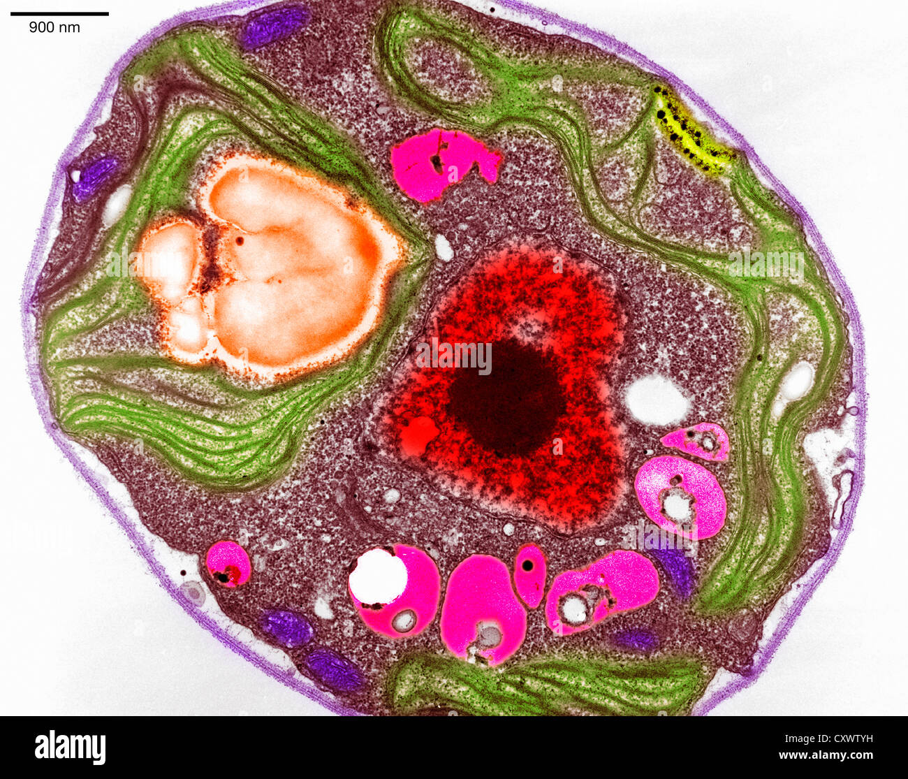
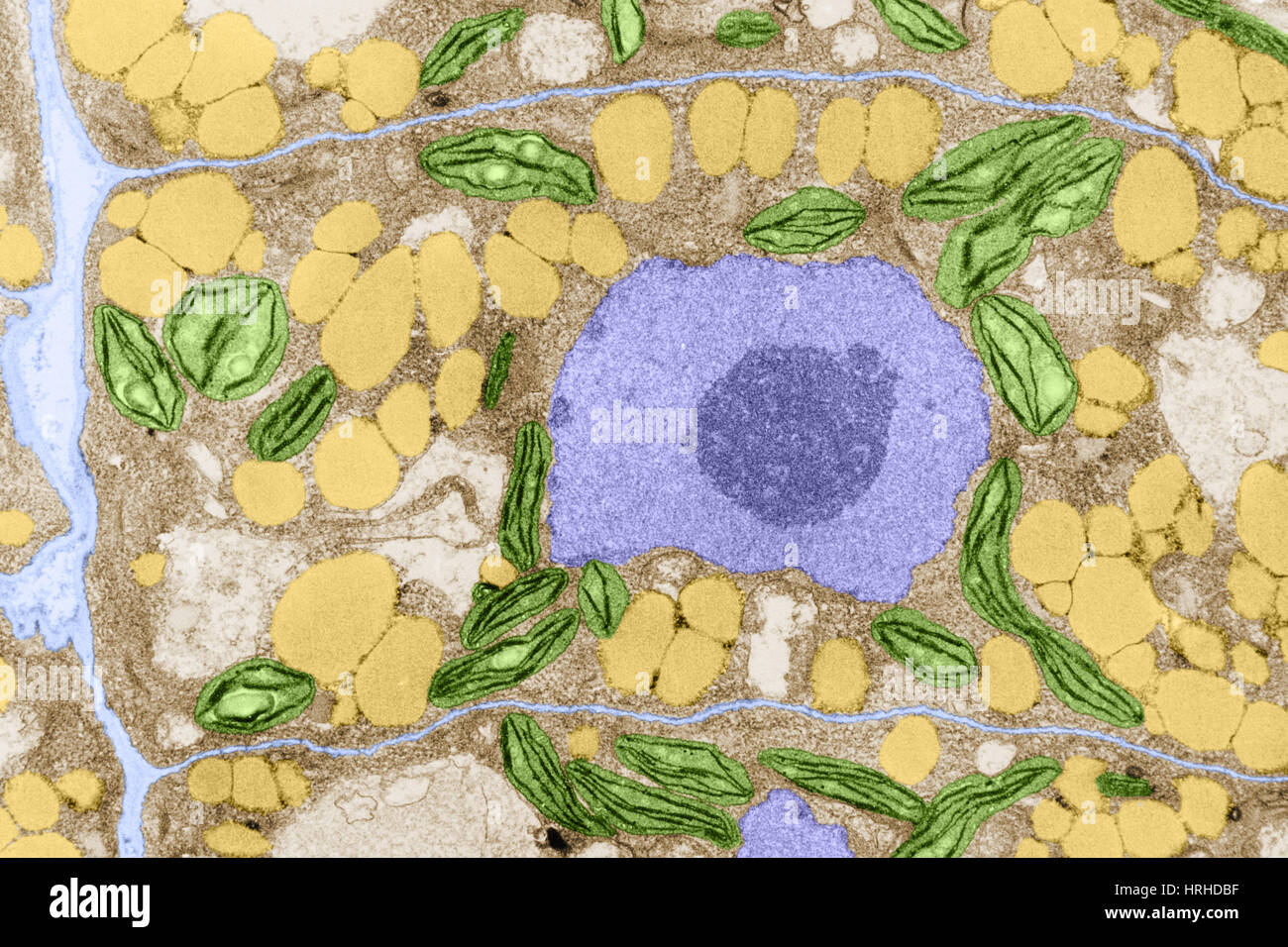
Plant cell tem hi-res stock photography and images - Alamy

Electron micrograph of cell walls in the cell vacuolization region just...  | Download Scientific Diagram
Electron micrograph of cell walls in the cell vacuolization region just... | Download Scientific Diagram

Cell surface and cell outline imaging in plant tissues using the  backscattered electron detector in a variable pressure scanning electron  microscope | Plant Methods | Full Text
Cell surface and cell outline imaging in plant tissues using the backscattered electron detector in a variable pressure scanning electron microscope | Plant Methods | Full Text

Image result for diagram of plant and animal cell under electron microscope  | Célula animal, Ciencias, Verdades absolutas
Image result for diagram of plant and animal cell under electron microscope | Célula animal, Ciencias, Verdades absolutas
Search in gallery
Search in gallery

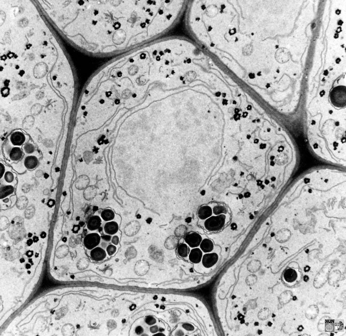
Cell Micrographs | BioNinja

Plant cell, TEM - Stock Image - C013/0796 - Science Photo Library

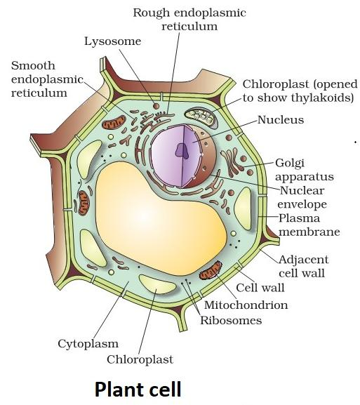
Illustrate only a plant cell seen under an electron microscope. How is it  different from animal cells?
Illustrate only a plant cell seen under an electron microscope. How is it different from animal cells?

Plant cell diagram, Cell theory, Cell diagram

Illustrate only a plant cell as seen under electron microscope. How is it  different from animal cell?
Illustrate only a plant cell as seen under electron microscope. How is it different from animal cell?
Plant_Bodies_Cells
Plant_Bodies_Cells

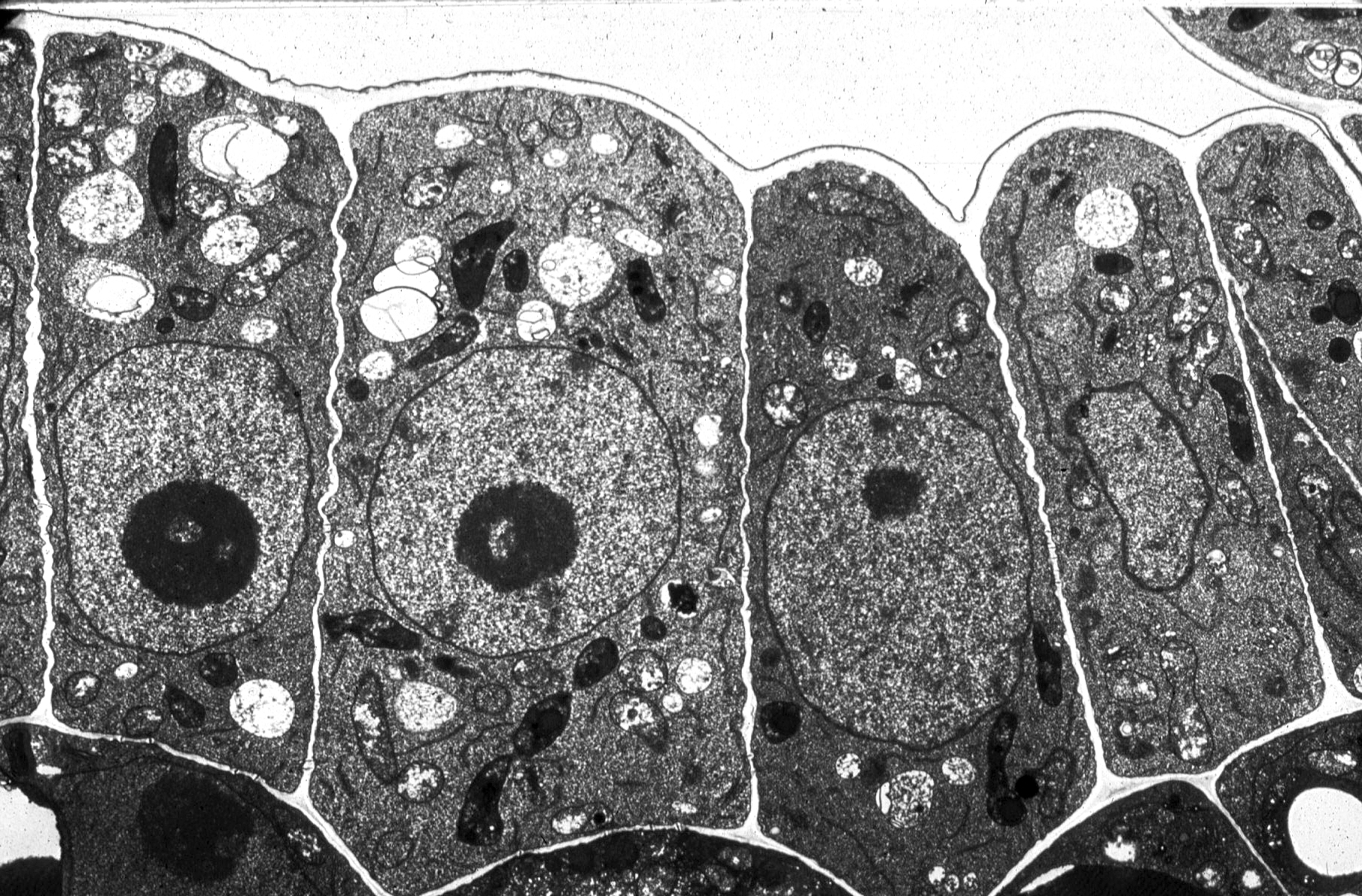
1.2 Skill: Interpretation of electron micrographs - YouTube

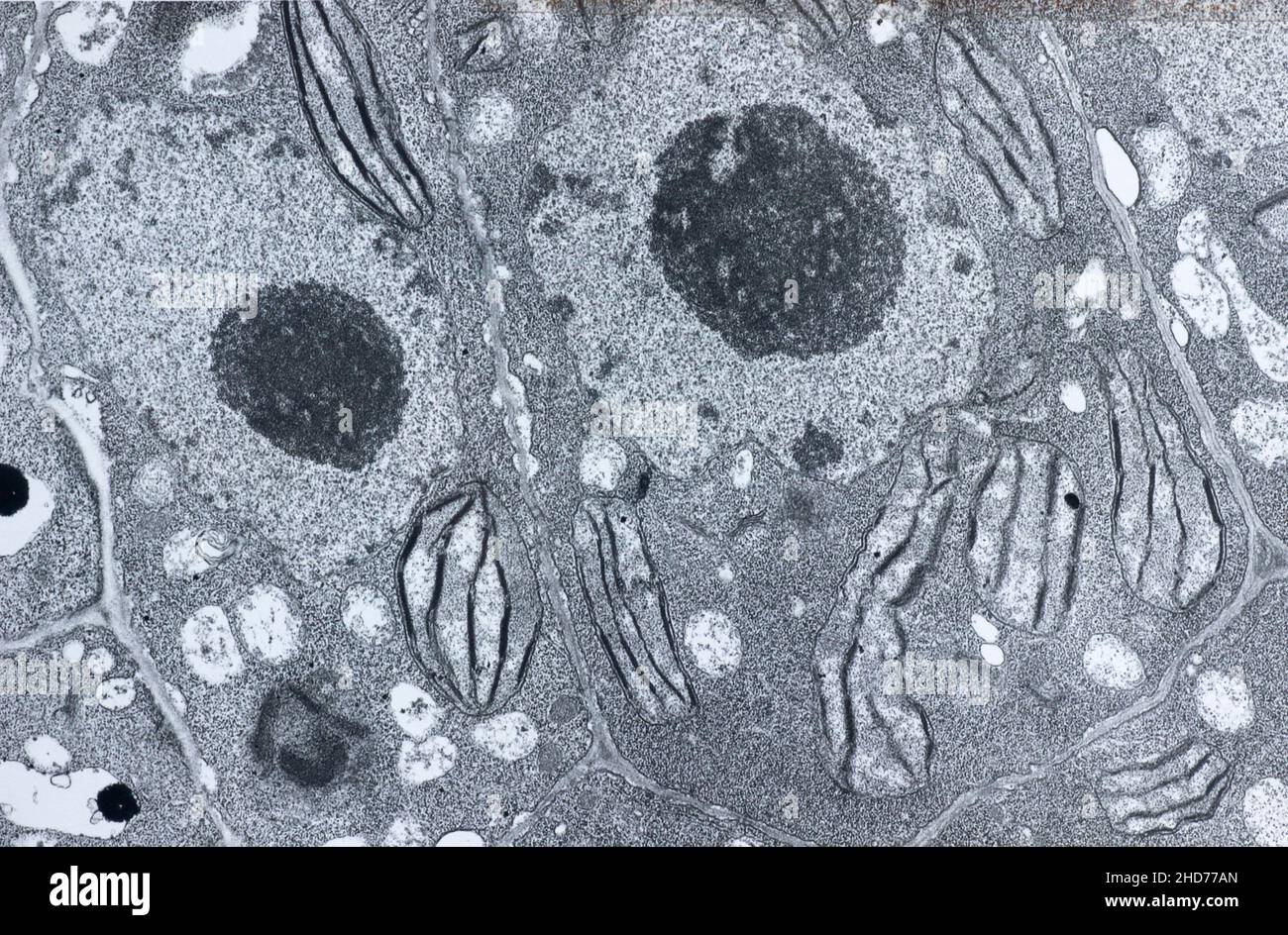
Electron micrograph plant cell hi-res stock photography and images - Alamy

Studies Using Electron Microscopy

Roger Innes: Faculty: About: Molecular Life Sciences Program: Indiana  University Bloomington
Roger Innes: Faculty: About: Molecular Life Sciences Program: Indiana University Bloomington

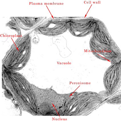
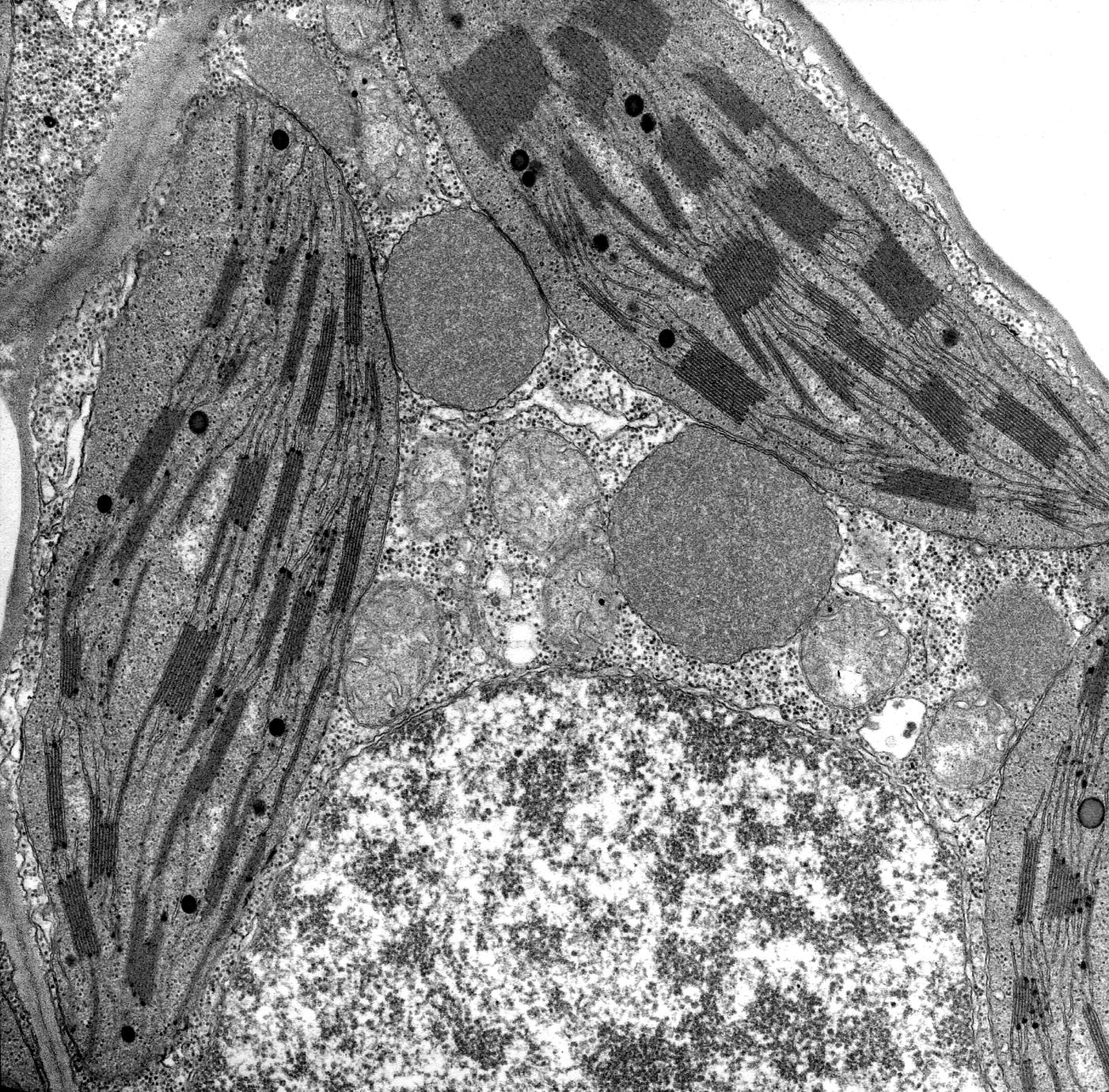
Nucleus, glyoxisomes, chloroplasts, and mitochondria - magnification at  13,900x - UWDC - UW-Madison Libraries
Nucleus, glyoxisomes, chloroplasts, and mitochondria - magnification at 13,900x - UWDC - UW-Madison Libraries
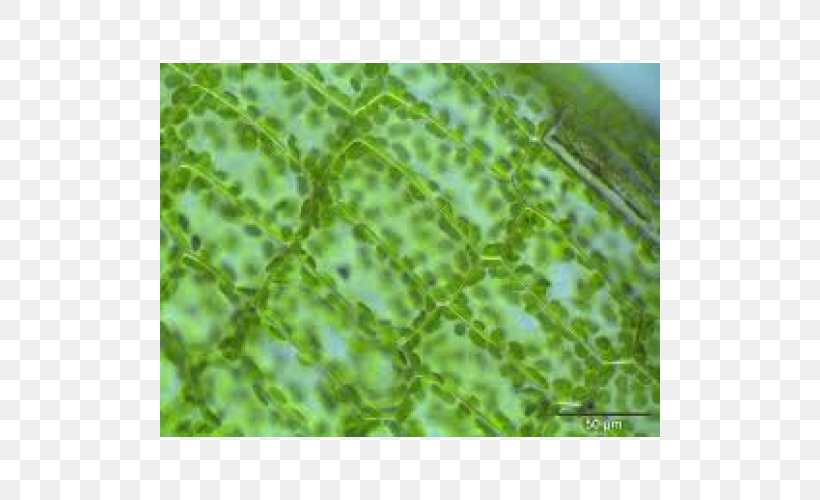
Light Plant Cell Microscope, PNG, 500x500px, Light, Biology, Cell, Electron  Microscope, Eukaryote Download Free
Light Plant Cell Microscope, PNG, 500x500px, Light, Biology, Cell, Electron Microscope, Eukaryote Download Free

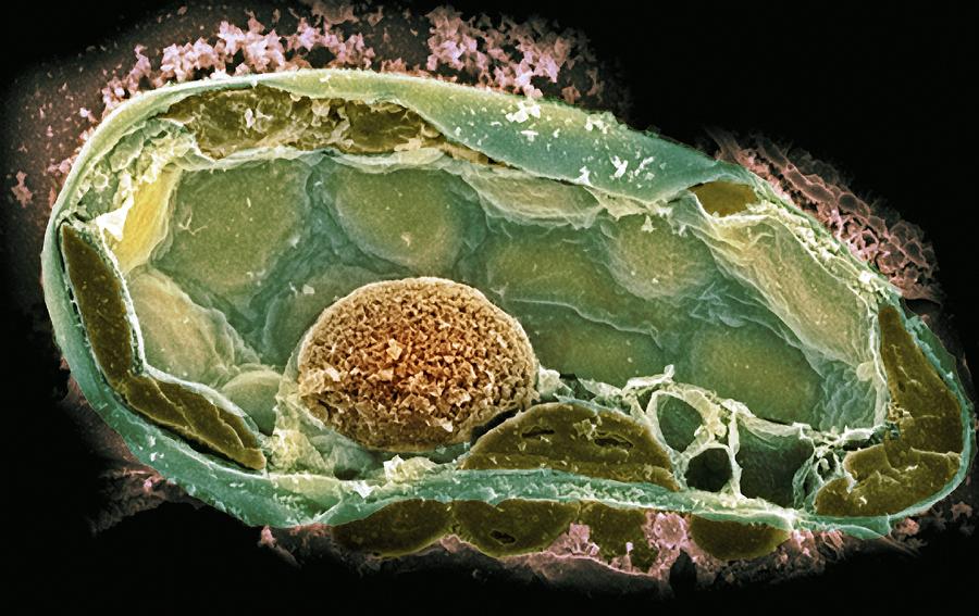
Plant Cell, Sem Photograph by Dr David Furness, Keele University - Pixels

Electron micrograph plant cell hi-res stock photography and images - Alamy

Biology 130 Lab 3 - Electron Micrographs

See flower cells in 3-D—no electron microscopy required

Electron micrograph showing the general appearance of a leaf cell from... |  Download Scientific Diagram
Electron micrograph showing the general appearance of a leaf cell from... | Download Scientific Diagram

Fast and cost-effective preparation of plant cells for scanning electron  microscopy (SEM) analysis - ScienceDirect
Fast and cost-effective preparation of plant cells for scanning electron microscopy (SEM) analysis - ScienceDirect